TL;DR:
AI is changing how bone fractures are detected by helping doctors spot injuries faster and more accurately. By analyzing X-rays, AI can catch small fractures that might be missed by the human eye, which is especially helpful for seniors and those with complex fractures.
AI doesn’t replace doctors; it supports them by providing extra insights and improving diagnosis speed. As AI tools become more common in clinics and hospitals, they’re making healthcare more reliable and efficient, ultimately leading to better outcomes for patients.
When someone gets hurt or takes a fall, an accurate diagnosis is the first step to recovery.
In hospitals and clinics around San Diego and across the country, radiology teams rely on X-rays to spot bone fractures. However, interpreting those images can be tough, even for skilled doctors. Small breaks or complex injuries may hide in plain sight, leading to missed diagnoses or delayed treatment.
In fact, missed fractures are estimated to account for up to 80% of diagnostic errors in the emergency department. This demonstrates just how frequently subtle injuries slip through initial evaluation.
Over the past few years, artificial intelligence (AI) has started to transform this process. New advances in medical imaging use smart computer systems to help radiologists catch fractures faster and more accurately.
For patients, this new era of diagnosis means a smoother path from X-ray to treatment, fewer errors, and possibly better health outcomes. This has real significance for seniors, Medicare beneficiaries, and families who want the best care with less risk of complications and more certainty in their recovery.
As more local hospitals and clinics consider adopting AI-based tools, there is growing interest in how these changes can support both providers and patients, especially in communities that value reliable, accessible healthcare. Let’s talk about it.
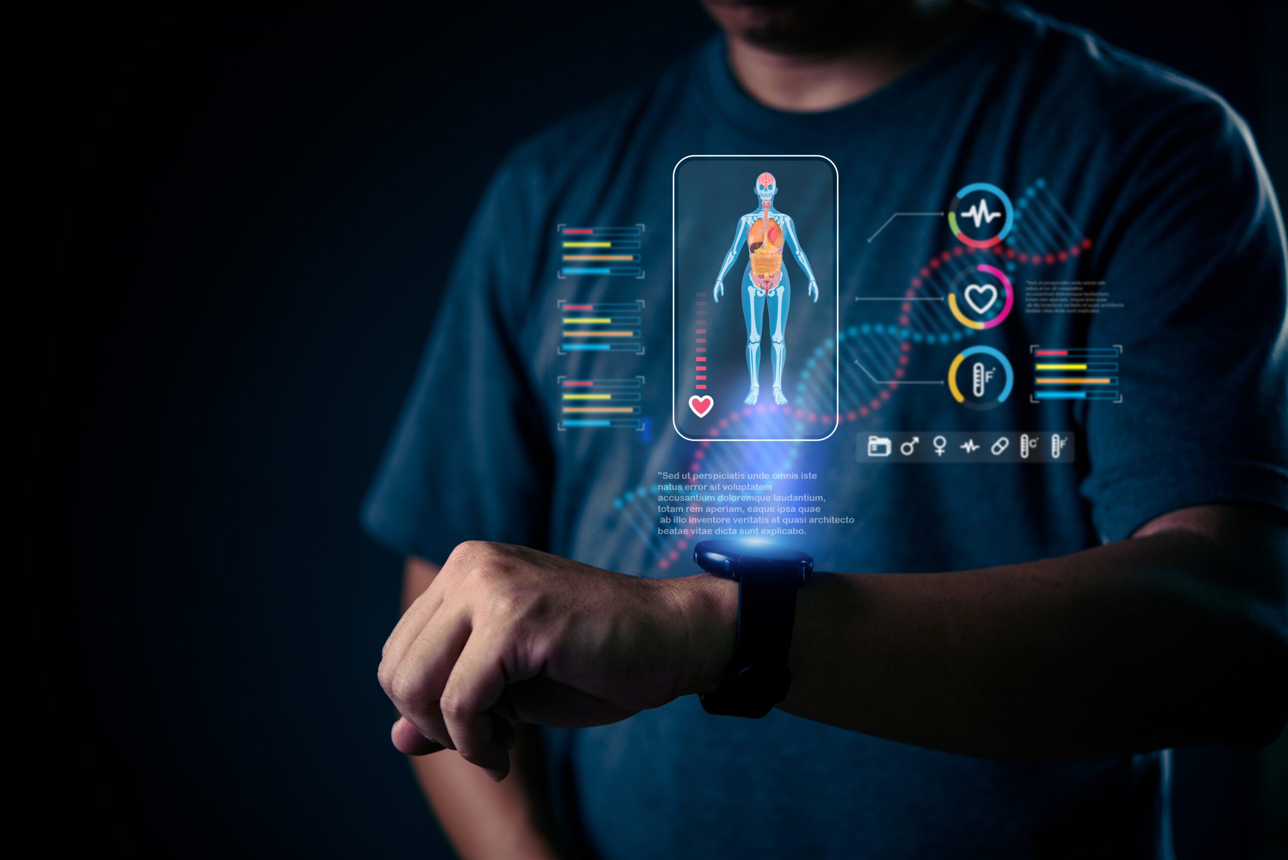
How AI Enhances Medical Imaging
Radiology relies heavily on X-rays and similar imaging tools to detect broken bones. Even with training, doctors may miss subtle evidence (hairline cracks or hidden damage), especially in a fast-paced setting.
Today, AI steps in as a digital partner that reviews these images with pinpoint accuracy.
Deep Learning Models in X-ray Analysis
AI algorithms rely on a technique known as “deep learning,” where computer systems learn from thousands of example images. These models analyze new X-rays, highlighting suspicious areas and flagging concerns that might otherwise go unnoticed. When a patient walks into an urgent care clinic in San Diego with a sore wrist or ankle, AI-assisted tools can quickly suggest if an image shows signs of a break.
Detecting Subtle Fractures Early
One of the greatest strengths of AI is its attention to detail across a huge volume of images. It can spot tiny cracks, slight misalignments, or faint shadowing that hints at a fresh break.
That early detection is vital for anyone (especially seniors) who might not recover well from a missed fracture.
Integrating with Existing Imaging Systems
Importantly, many AI solutions work hand in hand with the systems hospitals already use. Rather than replacing human clinicians, AI adds a “second set of eyes,” reducing the chances of oversight without slowing down care. Doctors still make the final call, but they do so with more information and support.
Improving Diagnostic Accuracy
While doctors and radiologists bring experience and clinical insight, research suggests even experts can miss subtle injuries, especially in busy emergency rooms. AI tools are designed to be tireless and consistent, which can improve both safety and speed.
Comparing AI and Human Performance
Recent studies show that, in some cases, AI matches or even slightly exceeds human accuracy in detecting fractures, particularly for certain bones like the wrist or hip.
In practical terms, this means fewer missed injuries and more reliable diagnoses for every patient.
AI-Based Fracture Detection for Subtle Injuries
Certain injuries, such as hairline wrist fractures or hip breaks in older adults, can be difficult to spot. AI aids by scanning thousands of pixels in every image, pointing out spots that need another look. This proactive approach can help doctors double-check and confirm their own findings.
Enhancing Radiology Reports
AI can also help draft clearer, more detailed radiology reports. These enhanced reports make life easier for the referring doctor, making sure the patient’s care plan is based on solid, comprehensive information.
AI Solutions for Complex Fracture Patterns
Not all fractures are simple. Some involve several bone pieces or unusual locations, requiring extra skill and attention to diagnose accurately. AI is playing a key role in helping providers handle these complex cases.
Below is a table highlighting how AI assists in the detection and classification of different fracture types, offering valuable insights that guide treatment decisions.
| Fracture Type | AI Assistance | Benefit for Providers |
| Proximal Humerus Fractures | AI classifies shoulder fractures using X-ray pattern recognition | Helps identify complex shoulder injuries and suggests treatment options |
| Wrist and Hip Fractures | AI analyzes the shape, size, and appearance of fractures | Reduces the risk of misdiagnosis, especially in elderly patients |
| Comprehensive Fracture Analysis | AI assesses fracture length, angle, and joint involvement | Supports tailored treatment plans, speeding up recovery for patients |
AI helps providers diagnose and treat fractures with more precision, reducing errors and enhancing the overall treatment process.
Now, let’s dive deeper into how AI specifically addresses these complex fracture types.
Classifying Proximal Humerus Fractures
Shoulder injuries, particularly near the top of the arm (proximal humerus), are common in older adults and can be complex. AI models now help classify these fractures by comparing X-ray patterns to large databases, suggesting the best treatment paths.
Wrist and Hip Fracture Detection
Wrist and hip breaks carry serious implications, especially for the elderly. AI can analyze the unique shapes and appearance of each fracture, helping providers avoid mistakes that could influence healing. For patients at local clinics or hospitals, this offers an extra layer of safety.
Comprehensive Fracture Pattern Analysis
Some AI systems go beyond simple detection, providing details about fracture length, angle, and possible involvement of nearby joints. This information can help orthopedic teams customize treatment plans, which is extremely beneficial for patients hoping to return to their daily routines quickly and safely.
Integration of AI in Clinical Practice
Bringing AI into real-world clinics and hospitals is an ongoing process. For best results, practices in San Diego must adapt workflows, ensure patient confidentiality, and build provider confidence with new tools.
Data Quality and Patient Data Privacy
Every AI system is only as good as the data it learns from. Local medical practices need robust policies to keep images and patient data private and secure, in keeping with California privacy laws and HIPAA regulations.
Training for Healthcare Providers
For AI to make a difference, doctors and radiologists need training in new systems. This includes understanding how to review AI findings, how to respond to alerts, and when to trust or question digital suggestions. Regular training ensures that technology benefits, rather than complicates, patient care.
Working with Existing Clinical Workflows
Integration works best when AI fits into the flow of care that most San Diego facilities already use. Many tools now connect directly to hospital imaging archives and electronic health records, allowing radiologists to access AI insights without delays or extra steps.
Impact on Patient Outcomes
Better diagnostic precision drives measurable gains in accuracy, treatment effectiveness, and overall quality of life for patients and their families. By strengthening clinical decision-making with reliable data, healthcare teams can respond sooner, choose the most effective interventions, and track real progress over time.
Early Detection Benefits for Seniors
For older adults, catching a fracture early means less risk of complications like infections, longer hospital stays, or loss of independence. AI speeds up the process, giving doctors more time to begin the right treatment, whether that means a cast, surgery, or physical therapy.
Reducing Missed Fractures
Each year, missed fractures account for a significant share of diagnostic errors in U.S. emergency rooms. AI, acting as a backup for the human eye, drives these numbers down, especially for hard-to-spot breaks.
Improved Patient Management and Follow-up
When diagnoses are more precise, follow-up care improves, too. Patients receive targeted recovery plans and better guidance on when to return for re-checks. For families coordinating care, especially across busy schedules, this means less uncertainty and more confidence in the process.
Challenges and Future Directions
Although AI has made impressive progress in detecting bone fractures, the field is still maturing. Recognizing both its limitations and potential helps clinicians and patients maintain realistic expectations as adoption grows.
Deep Learning Algorithm Limitations
AI tools depend heavily on high-quality, diverse data and continuous validation. A 2024 systematic review in Radiology found pooled sensitivity and specificity around 92% for fracture detection (comparable to human readers), but most studies lacked external validation and real-world testing. Models trained on narrow datasets can misinterpret images from different hospitals or patient groups, and their accuracy declines when imaging protocols or equipment change.
Pediatric cases remain especially difficult, where variable bone development and smaller datasets challenge model reliability. For now, AI will likely complement, not replace, clinical judgment.
Continuous Improvement of AI Models
Researchers worldwide, including leading centers in California, are working to make diagnostic AI smarter, fairer, and more generalizable. Current efforts focus on increasing dataset diversity, reducing algorithmic bias, and embedding continuous feedback from clinicians.
As these models evolve, the goal is not just better accuracy but dependable performance across varied healthcare settings.
Expanding Applications Beyond Bone Fractures
Fracture detection is only the beginning. Similar deep-learning approaches are already being tested for cancer screening, cardiovascular imaging, and neurological conditions.
Studies published in Frontiers in Radiology and JAMA Network Open highlight promising early results, suggesting that the same algorithms used for bone imaging could eventually support diagnosis and triage across multiple organ systems.
As the technology advances, local hospitals and clinics may see AI integrated into routine imaging workflows, speeding up diagnosis, prioritizing urgent cases, and expanding access to expert-level analysis.
The Future of Fracture Care Is Getting Clearer
Across the nation, advances in medical imaging are helping doctors work faster, diagnose with greater precision, and spend more time focused on patients, not paperwork. AI in radiology isn’t replacing expertise, but rather refining it to give healthcare teams sharper tools to deliver accurate, timely answers.
For Medicare beneficiaries and families who count on dependable coverage, that progress matters. As technology evolves, having a great insurance partner helps you stay prepared for new options in care and diagnostics.
If you’d like to understand how these innovations may affect your Medicare choices or medical coverage, reach out to Terri Yurek Insurance today. We’re here to help you make sense of what’s next in healthcare.
Frequently Asked Questions (FAQs)
1. What exactly is AI in radiology?
- AI in radiology uses smart computer algorithms (like deep learning) to analyze medical images, such as X-rays. It helps radiologists spot issues, like bone fractures, more quickly and accurately than they might on their own. Think of it as a digital assistant that gives doctors an extra set of eyes.
2. How does AI actually help in finding bone fractures?
- AI scans X-ray images and looks for any signs of fractures, including those tiny cracks or hidden breaks that could be easy to miss. It highlights these areas, making sure the doctor doesn’t overlook anything important, leading to a more precise diagnosis.
3. What are the main benefits of using AI in healthcare?
- AI helps doctors catch fractures faster and more accurately, reduces the chances of missed diagnoses, and speeds up the entire process. This means patients, especially older adults, get the treatment they need without unnecessary delays or errors, ultimately improving their recovery.
4. Can AI replace radiologists?
- No, AI isn’t here to replace radiologists. It’s there to support them. Radiologists still make the final call, but AI assists by providing valuable insights that make their job easier and more efficient. It’s all about working together to improve patient care.
5. Are there any limitations to AI in radiology?
- Yes, AI works best when it’s trained on high-quality data, and it might struggle with certain cases, like those involving younger patients or unusual imaging conditions. It also needs continuous updates and validation to ensure it stays reliable. Right now, AI is a tool to help doctors, not a one-size-fits-all solution.